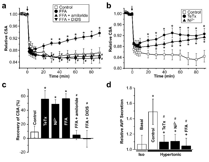
Fig. 3. The effects of FFA, TeTx and Ni2+ on osmotic shrinkage in and AVP secretion from AVP neurons in response to hyperosmotic stimulation. (a) Time course of changes in the cross-sectional area (CSA) of the cell soma before and after application of hyperosmotic bath solution (at arrow) in the absence (Control) or presence of FFA (100 mM), FFA plus amiloride (100 mM) or FFA plus DIDS (100 mM) (n=11-19). (b) Time course of changes in the cross-sectional area (CSA) of the cell soma before and after application of hyperosmotic bath solution (at arrow) in the absence (Control) or presence of TeTx (15 nM) or Ni2+ (3 mM) (n=8-14). TeTx was applied for 70 min before hyperosmotic stimulation as well. (c) Percent recovery of CSA (from the average of Control peak osmotic shrinking) at 90 min after a hyperosmotic challenge in the absence or presence of drugs (n = 9-19). *p<0.05 vs. Control. #p<0.05 vs. in the presence of FFA. (d) The relative amount of AVP released from dissociated SON neurons for 90 min after application of isosmotic (Basal; n=16) or hyperosmotic bath solution in the absence (Control) or presence of TeTx, Ni2+, or FFA (n=7-21). *p<0.05 vs. Basal. #p<0.05 vs. Control.